Advanced Cardiac Imaging
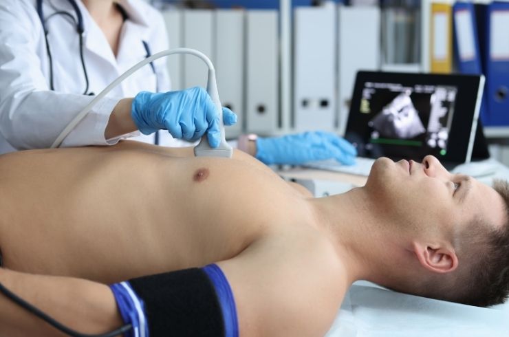
Advanced Cardiac Imaging Services for Detailed Heart Evaluation in Amravati, Maharashtra
Advanced cardiac imaging uses specialized techniques to evaluate the heart’s structure, function, blood flow and movement. Dr. Niraj provides cardiac imaging assessment in Amravati, Maharashtra, with the appropriate imaging approach selected according to the patient’s symptoms, clinical condition and diagnostic requirements.
What it is
Advanced cardiac imaging includes different echocardiographic techniques that provide detailed information about the heart.
These may include:
- 2D Echocardiography
- Doppler
- 3D / 4D Echocardiography
- TEE
- Stress Echocardiography
- Strain Imaging
- Congenital Echocardiography
- Structural Heart Imaging
- Pre- and post-intervention imaging
The appropriate imaging technique depends on the clinical question and technology available.
2D Echocardiography
2D Echocardiography uses ultrasound to create real-time images of the heart.
It can help assess:
- Heart chambers
- Heart muscle movement
- Pumping function
- Heart valves
- Structural abnormalities
- Pericardial fluid
It is a non-invasive examination and does not use ionizing radiation.
Doppler
Doppler echocardiography evaluates the movement of blood through the heart and blood vessels.
It can help assess:
- Blood-flow direction
- Blood-flow velocity
- Valve function
- Abnormal blood flow
- Pressure-related information
Doppler assessment is commonly combined with echocardiography when evaluating cardiac function and valve disease.
3D / 4D Echocardiography
3D / 4D echocardiography provides three-dimensional visualization of cardiac structures, with 4D imaging incorporating movement over time.
It may provide additional anatomical information in selected situations, particularly when detailed assessment of cardiac structures or valves is required.
Its usefulness depends on image quality, the clinical question and the technology available.
Transoesophageal Echocardiography (TEE)
Transoesophageal Echocardiography (TEE) uses an ultrasound probe positioned in the oesophagus to obtain detailed images of the heart.
Because the oesophagus lies close to the heart, TEE can provide detailed visualization of selected cardiac structures when standard transthoracic imaging does not provide sufficient information.
TEE is an invasive examination and requires appropriate preparation and clinical supervision.
Stress Echocardiography
Stress Echocardiography combines cardiac ultrasound imaging with controlled cardiac stress to evaluate how the heart responds.
It may be used in selected patients to assess:
- Heart muscle movement
- Functional response to stress
- Possible evidence of reduced blood supply
- Exercise or stress-related cardiac symptoms
The type of stress used depends on the patient’s clinical circumstances.
Strain Imaging
Strain imaging provides additional information about how different areas of the heart muscle deform and contract.
It can help provide more detailed assessment of myocardial function beyond conventional measurements in selected clinical situations.
Strain assessment may be useful when evaluating cardiac function in appropriate patients.
Congenital Echocardiography
Congenital echocardiography focuses on assessing structural heart abnormalities that are present from birth.
It may help evaluate conditions involving:
- Heart chambers
- Septal defects
- Valves
- Major blood vessels
- Abnormal blood flow
- Other congenital cardiac structures
The imaging approach depends on the patient’s age, anatomy and suspected congenital condition.
Structural Heart Imaging
Structural heart imaging supports the assessment of conditions affecting the heart’s valves and other structural components.
It may contribute to evaluation and planning for selected:
- Aortic valve conditions
- Mitral valve conditions
- Tricuspid valve conditions
- Congenital structural abnormalities
- Catheter-based structural interventions
Detailed imaging may be important for patient selection and procedural planning.
Pre-Intervention Imaging
Imaging performed before an intervention can help establish the relevant cardiac anatomy and provide information needed for treatment planning.
Depending on the procedure, imaging may assess:
- Valve anatomy
- Heart chambers
- Blood flow
- Structural abnormalities
- Cardiac function
- Relevant measurements
The exact imaging requirements depend on the planned intervention.
Post-Intervention Imaging
Imaging after a cardiac intervention can help evaluate the result of treatment.
It may be used to assess:
- Heart function
- Valve function
- Blood flow
- Device or intervention-related findings
- Residual abnormalities
- Changes following treatment
Follow-up imaging is planned according to the patient’s condition and the procedure performed.
Who May Need Advanced Cardiac Imaging
Advanced cardiac imaging may be considered for patients with:
- Breathlessness
- Chest discomfort
- Heart murmurs
- Palpitations
- Heart failure
- Cardiomyopathy
- Heart valve disease
- Congenital heart disease
- Suspected structural heart disease
- Known cardiac disease requiring monitoring
- Patients undergoing or recovering from selected cardiac interventions
Causes and Conditions Assessed
Advanced cardiac imaging can contribute to the evaluation of:
- Coronary artery disease
- Heart failure
- Cardiomyopathy
- Heart valve disease
- Structural heart disease
- Congenital heart disease
- Abnormal heart function
- Selected post-intervention conditions
Imaging findings are interpreted together with symptoms, examination findings and other investigations.
Risk Factors
Patients with cardiovascular risk factors or known cardiac conditions may require cardiac imaging based on their clinical presentation.
Relevant factors may include:
- High blood pressure
- Diabetes
- High cholesterol
- Coronary artery disease
- Previous heart attack
- Heart failure
- Cardiomyopathy
- Heart valve disease
- Congenital heart disease
- Abnormal heart rhythms
Having a risk factor does not automatically mean advanced cardiac imaging is required.
Symptoms That May Require Evaluation
Cardiac imaging may be considered when patients experience:
- Breathlessness
- Chest discomfort
- Palpitations
- Fatigue
- Dizziness
- Fainting
- Reduced exercise tolerance
- Leg or ankle swelling
The need for imaging depends on the overall clinical assessment.
Red-Flag Symptoms
Advanced cardiac imaging should not delay emergency care.
Seek immediate emergency medical attention for:
- Severe or persistent chest pain
- Severe breathlessness
- Fainting or loss of consciousness
- Sudden severe weakness
- New neurological symptoms
- Symptoms suggestive of a heart attack
Diagnosis
Advanced cardiac imaging may contribute to the diagnosis and assessment of:
- Heart failure
- Cardiomyopathy
- Valve disease
- Structural heart disease
- Congenital heart disease
- Abnormal cardiac function
- Selected coronary conditions
Additional investigations may be required depending on the suspected diagnosis.
When Advanced Cardiac Imaging May Be Required
Advanced imaging may be recommended when detailed information is needed about:
- Heart function
- Valve structure and function
- Blood flow
- Congenital cardiac anatomy
- Structural heart disease
- Myocardial function
- Pre-intervention anatomy
- Post-intervention results
The most appropriate imaging technique is selected according to the clinical question.
Medical Treatment
Advanced cardiac imaging is a diagnostic and monitoring service rather than a treatment itself.
The findings may help guide treatment for:
- Heart failure
- Cardiomyopathy
- Valve disease
- Structural heart disease
- Congenital heart disease
- Other cardiac conditions
Treatment decisions depend on the complete clinical assessment.
Treatment Alternatives
Depending on the diagnostic question, other investigations may include:
- ECG
- Holter monitoring
- 2D Echocardiography
- CT cardiac imaging
- CT coronary angiography
- Cardiac MRI
- Coronary angiography
- Other specialized cardiac investigations
The appropriate investigation depends on the patient’s symptoms and suspected condition.
Related Procedures
Related cardiac services include:
- 2D Echocardiography
- Doppler
- 3D / 4D Echocardiography
- TEE
- Stress Echocardiography
- Strain Imaging
- Congenital Echocardiography
- Structural Heart Imaging
- Coronary Angiography
- Intravascular Imaging
- Structural Heart Programme
- TAVR / TAVI
- Mitral Valve Interventions
- Tricuspid Interventions
FAQs
What is advanced cardiac imaging?
Advanced cardiac imaging uses specialized imaging techniques to evaluate the heart’s structure, function, blood flow and movement in greater detail.
What is the difference between 2D and 3D / 4D echocardiography?
2D echocardiography produces two-dimensional real-time images, while 3D / 4D echocardiography can provide three-dimensional visualization and movement over time in suitable clinical situations.
What is Doppler echocardiography?
Doppler echocardiography evaluates blood flow through the heart and vessels and can provide information about blood-flow direction and velocity.
What is TEE?
TEE, or transoesophageal echocardiography, uses an ultrasound probe positioned in the oesophagus to obtain detailed images of selected cardiac structures.
What is stress echocardiography?
Stress echocardiography evaluates how the heart muscle responds to controlled stress and can help assess selected patients for possible reduced blood supply or other functional abnormalities.
What is strain imaging?
Strain imaging provides additional information about how different regions of the heart muscle contract and deform.
What is congenital echocardiography?
Congenital echocardiography evaluates the structure and function of the heart in patients with suspected or known congenital heart abnormalities.
Why is cardiac imaging performed before an intervention?
Pre-intervention imaging can provide important information about cardiac anatomy, function and measurements needed for treatment planning.
Why is imaging performed after a cardiac intervention?
Post-intervention imaging can help assess heart function, blood flow, valve function and findings related to the completed procedure.
Does echocardiography use radiation?
No. Echocardiography uses ultrasound waves and does not use ionizing radiation.
When should I consult Dr. Niraj in Amravati?
Patients with breathlessness, chest discomfort, palpitations, heart murmurs, heart failure, valve disease, congenital heart disease or other cardiac concerns can consult Dr. Niraj to determine whether cardiac imaging is appropriate.
Book an Appointment with Dr. Niraj for Advanced Cardiac Imaging in Amravati
If you need 2D Echo, Doppler, 3D / 4D Echo, TEE, stress echocardiography, strain imaging or structural heart imaging, Dr. Niraj can evaluate your clinical requirements and determine the appropriate cardiac imaging pathway in Amravati, Maharashtra.