Intravascular Imaging
Intravascular Imaging for Advanced PCI Assessment and Optimisation in Amravati, Maharashtra
Intravascular imaging is an advanced technique used during selected coronary interventions to obtain detailed information from inside the coronary artery. It can provide information about vessel dimensions, plaque, calcium and stent placement that may not be fully visible through conventional angiography.
For patients seeking intravascular imaging and advanced PCI evaluation in Amravati, Maharashtra, Dr. Niraj can assess whether intravascular imaging may add useful information to coronary diagnosis, procedural planning or stent optimisation.
What it is
Intravascular imaging uses a specialized catheter-based imaging system positioned inside a coronary artery.
The information obtained can help assess:
- Vessel dimensions
- Plaque burden
- Calcium
- Lesion characteristics
- Stent sizing
- Stent expansion
- Stent apposition
- Stent edges
- Left-main coronary anatomy
- Overall PCI result
The use of intravascular imaging depends on the patient’s anatomy, clinical situation and procedural requirements.
IVUS Principles
Intravascular ultrasound (IVUS) uses ultrasound technology to create cross-sectional images from inside the coronary artery.
IVUS can provide information about the vessel wall and lumen and may help the interventional team understand coronary anatomy more precisely during selected procedures.
It can be particularly useful in complex coronary lesions where angiography alone may not provide sufficient information for treatment planning or optimisation.
Vessel Dimensions
Accurate assessment of vessel dimensions can help with appropriate device and stent selection.
IVUS may provide information about:
- Vessel diameter
- Reference vessel dimensions
- Lumen dimensions
- Vessel morphology
- Lesion location
This information can support more individualized PCI planning.
Plaque Burden
Intravascular imaging can help visualize plaque within the coronary artery and assess its distribution.
Understanding plaque burden and lesion characteristics may assist in:
- Assessing lesion complexity
- Planning PCI
- Selecting an appropriate treatment strategy
- Understanding the coronary anatomy beyond the angiographic appearance
Calcium Assessment
IVUS can help identify and assess coronary calcium.
Calcium assessment may be particularly important when planning PCI for heavily calcified lesions because severe calcium can make balloon expansion and stent deployment more challenging.
In selected cases, calcium assessment can help determine whether additional lesion-preparation techniques may be required.
Stent Sizing
Appropriate stent sizing is an important part of PCI.
Intravascular imaging may help assess the dimensions of the treated vessel and reference segments, supporting selection of an appropriate stent size.
This can be particularly useful in complex coronary anatomy where accurate vessel measurement is important.
Stent Expansion
After stent implantation, intravascular imaging can help evaluate whether the stent has expanded appropriately.
Assessment may include:
- Stent expansion
- Stent symmetry
- Residual narrowing
- Relationship between the stent and vessel wall
In selected cases, this information can identify areas that may require further optimisation.
Stent Apposition
Stent apposition refers to how well the stent is positioned against the vessel wall.
Intravascular imaging can help identify areas where the stent may not be adequately opposed to the vessel wall.
This can provide additional information for optimisation following PCI.
Edge Problems
Intravascular imaging can help assess the areas immediately before and after a coronary stent.
It may help identify:
- Residual plaque
- Significant narrowing
- Dissection
- Other abnormalities near the stent edges
This information can be useful when evaluating the final result of selected complex PCI procedures.
Left-Main Assessment
Left-main coronary disease requires careful anatomical assessment because the left main artery supplies a substantial portion of the heart muscle.
IVUS may provide detailed information about:
- Left-main vessel dimensions
- Lumen characteristics
- Plaque distribution
- Lesion severity
- Stent expansion following left-main PCI
Its use depends on the individual clinical and procedural circumstances.
PCI Optimisation
Intravascular imaging can be used at different stages of selected PCI procedures.
It may assist with:
- Pre-procedure lesion assessment
- Vessel sizing
- Stent selection
- Stent deployment
- Assessment of stent expansion
- Assessment of stent apposition
- Identification of edge problems
- Final PCI optimisation
This can be especially valuable in complex coronary interventions where precise anatomical information is important.
OCT May Be Included Only If Clinically Offered
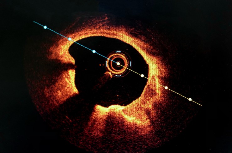
Optical coherence tomography (OCT) is another form of intravascular imaging that uses light-based technology to produce high-resolution images inside the coronary artery.
OCT may be considered only where it is clinically offered and appropriate.
It should not be presented as routinely available unless it is specifically offered as part of the patient’s care.
Causes
Intravascular imaging itself is not a treatment for a particular disease. It is an imaging technique used to evaluate coronary artery disease and assist selected PCI procedures.
Conditions where it may be relevant include:
- Complex coronary artery disease
- Calcified coronary lesions
- Left-main coronary disease
- Bifurcation disease
- In-stent restenosis
- Complex PCI
- Uncertain coronary anatomy requiring additional assessment
Risk Factors
The underlying coronary conditions that may lead to a need for advanced coronary assessment are associated with risk factors such as:
- Diabetes
- High blood pressure
- High cholesterol
- Smoking or tobacco use
- Obesity
- Physical inactivity
- Increasing age
- Family history of coronary artery disease
- Chronic kidney disease
- Previous coronary disease
Symptoms
Symptoms depend on the underlying coronary condition rather than the imaging procedure itself.
Possible symptoms of coronary disease include:
- Chest discomfort
- Breathlessness during activity
- Reduced exercise tolerance
- Unusual fatigue
- Sweating
- Dizziness
- Discomfort spreading to the arm, back, neck or jaw
Some patients may have significant coronary disease without typical symptoms.
Red-Flag Symptoms
Seek immediate emergency medical attention for:
- Severe or persistent chest pain
- Chest discomfort occurring at rest
- Sudden severe breathlessness
- Fainting
- Cold sweating with chest discomfort
- Severe weakness
- New confusion
- Pain spreading to the arm, back, neck or jaw
These symptoms may indicate an acute cardiac emergency.
Diagnosis
Intravascular imaging is generally used as an additional assessment tool rather than as a standalone diagnostic test.
Depending on the clinical situation, evaluation may include:
- Medical history
- Physical examination
- ECG
- Blood tests
- Echocardiography
- Non-invasive cardiac testing
- Coronary CT angiography where appropriate
- Coronary angiography
- Intravascular imaging
The need for IVUS or another imaging technique depends on the clinical question and coronary anatomy.
When Coronary Angiography May Be Required
Coronary angiography may be performed when detailed visualization of the coronary arteries is required.
It may be considered for:
- Suspected significant coronary disease
- Persistent or worsening angina
- Acute coronary syndrome
- Complex coronary anatomy
- Planning PCI
- Assessment of previously treated coronary lesions
Intravascular imaging may then provide additional information during selected procedures.
Medical Treatment
Medical treatment for underlying coronary artery disease may include:
- Cholesterol-lowering medicines
- Antiplatelet medicines when clinically indicated
- Anti-anginal medicines
- Blood-pressure management
- Diabetes management
- Smoking cessation
- Weight management
- Heart-healthy dietary changes
- Appropriate physical activity
- Cardiac rehabilitation where suitable
The appropriate treatment depends on the patient’s individual condition.
When Intervention May Be Required
PCI may be considered when a coronary lesion is clinically significant and revascularization is appropriate.
Intravascular imaging may be particularly useful during selected complex interventions involving:
- Left-main disease
- Calcified lesions
- Bifurcation lesions
- Complex coronary anatomy
- In-stent restenosis
- Other lesions where angiography alone may not provide sufficient information
Treatment Alternatives
Depending on the underlying coronary disease, treatment options may include:
- Medical therapy
- Lifestyle and risk-factor management
- PCI
- CABG
- Further diagnostic assessment
- Heart Team evaluation for complex coronary disease
The appropriate strategy depends on symptoms, coronary anatomy, heart function and overall cardiovascular risk.
Related Procedures
Related procedures include:
- Coronary Angiography
- Complex PCI
- Left Main PCI
- Bifurcation PCI
- CTO Angioplasty
- Calcified Lesion PCI
- Rotablation
- Intravascular Lithotripsy (IVL)
- Coronary Physiology
- Multivessel PCI
- Coronary Artery Bypass Grafting (CABG)
FAQs
What is intravascular imaging?
Intravascular imaging is a catheter-based technique that provides detailed images from inside a coronary artery during selected cardiac evaluations and PCI procedures.
What is IVUS?
IVUS, or intravascular ultrasound, uses ultrasound technology to create cross-sectional images of the coronary artery from inside the vessel.
How does IVUS help during PCI?
IVUS can help assess vessel dimensions, plaque, calcium, stent sizing, stent expansion, apposition and other aspects of the PCI result.
Can IVUS assess coronary calcium?
Yes. IVUS can provide information about the presence and characteristics of calcium within selected coronary lesions.
Can IVUS help with left-main PCI?
Yes. IVUS may provide detailed information about left-main anatomy, vessel dimensions and stent expansion during selected left-main PCI procedures.
Why is stent expansion assessed?
Assessing stent expansion can help identify whether the implanted stent has achieved an appropriate result and whether further optimisation may be required.
What is stent apposition?
Stent apposition describes how well the stent is positioned against the vessel wall. Intravascular imaging can help identify areas of incomplete apposition.
Is OCT the same as IVUS?
No. IVUS uses ultrasound, while OCT uses light-based imaging. OCT may be used only where it is clinically offered and appropriate.
Does every PCI require intravascular imaging?
No. Intravascular imaging is used selectively depending on the patient’s coronary anatomy, clinical situation and procedural requirements.
When should I consult Dr. Niraj in Amravati?
Patients with complex coronary disease, calcified lesions, left-main disease, bifurcation disease or challenging PCI planning may benefit from specialist assessment by Dr. Niraj.
Book an Appointment with Dr. Niraj for Intravascular Imaging Evaluation in Amravati
If you have been diagnosed with complex coronary disease or have been advised to undergo advanced PCI, Dr. Niraj can assess your coronary anatomy and determine whether intravascular imaging may provide useful information for treatment planning or PCI optimisation.