Calcified Coronary Lesions
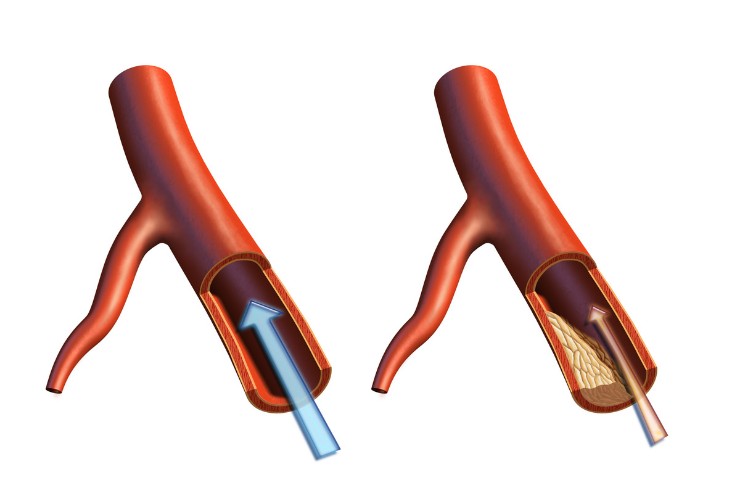
Calcified Coronary Lesions Treatment and Advanced PCI Care in Amravati, Maharashtra
Calcified coronary lesions occur when calcium builds up within the walls of the coronary arteries, making the affected vessel more rigid and sometimes more difficult to treat with conventional angioplasty and stent placement.
For patients seeking calcified coronary lesion treatment in Amravati, Maharashtra, Dr. Niraj evaluates the extent and characteristics of coronary calcium and selects an appropriate lesion-preparation strategy based on coronary anatomy, calcium severity, symptoms, and overall cardiovascular condition.
What it is
Calcified coronary lesions are coronary artery blockages containing significant calcium deposits within the arterial wall.
Calcium can make a coronary artery less flexible and may interfere with balloon expansion and proper stent deployment. Advanced assessment and lesion-preparation techniques may therefore be required before stent implantation.
Treatment strategies may include:
- IVUS
- Rotational atherectomy
- Intravascular lithotripsy
- Cutting/scoring balloons where applicable
- Lesion preparation
- Stent expansion and optimisation
- Combined strategies where appropriate
Causes
Coronary calcification generally develops as part of the long-term atherosclerotic process.
As coronary plaque develops over time, calcium may accumulate within the arterial wall. More advanced or long-standing coronary artery disease may be associated with heavier calcification.
Calcification can also be encountered in patients with extensive coronary disease and certain chronic medical conditions.
Risk factors
Factors associated with coronary calcification and coronary artery disease include:
- Increasing age
- Diabetes
- High blood pressure
- High cholesterol
- Smoking or tobacco use
- Chronic kidney disease
- Obesity
- Physical inactivity
- Family history of coronary artery disease
- Long-standing coronary artery disease
- Previous coronary interventions
The presence of multiple risk factors can contribute to progressive coronary plaque and calcification.
Symptoms
Calcified coronary lesions may produce symptoms similar to other forms of coronary artery disease.
Possible symptoms include:
- Chest pressure or discomfort
- Breathlessness during physical activity
- Reduced exercise tolerance
- Unusual fatigue
- Dizziness
- Sweating
- Discomfort spreading to the arm, shoulder, back, neck or jaw
Some patients may have significant coronary disease without typical symptoms.
Red-flag symptoms
Seek immediate emergency medical attention if you experience:
- Severe or persistent chest pain
- New chest pressure occurring at rest
- Sudden severe breathlessness
- Cold sweating with chest discomfort
- Fainting
- Severe weakness
- New confusion
- Pain spreading to the arm, back, neck or jaw
- Rapid worsening of symptoms in a patient with known coronary disease
These symptoms may indicate acute coronary syndrome or a heart attack.
Diagnosis
Assessment of calcified coronary disease involves understanding both the degree of narrowing and the amount and distribution of calcium.
Evaluation may include:
- Medical history
- Physical examination
- ECG
- Blood tests
- Echocardiography
- Coronary CT imaging where appropriate
- Coronary angiography
- Intravascular ultrasound (IVUS)
- Other intravascular imaging techniques where appropriate
Angiography can demonstrate the coronary anatomy, while intravascular imaging can provide additional information about the vessel wall and calcium characteristics.
When coronary angiography may be required
Coronary angiography may be recommended when significant coronary disease is suspected or when detailed assessment is needed before an intervention.
It may be considered for patients with:
- Persistent or worsening angina
- Abnormal cardiac investigations
- Suspected significant coronary artery disease
- Known heavily calcified coronary lesions
- Complex coronary anatomy
- Acute coronary syndrome
- Need for PCI planning
Angiography helps determine the location and severity of coronary narrowing and assists in planning appropriate intervention.
Medical treatment
Medical treatment remains an important part of managing coronary artery disease, including calcified lesions.
Depending on the patient’s individual condition, treatment may include:
- Cholesterol-lowering medicines
- Antiplatelet medicines when clinically indicated
- Anti-anginal medicines
- Blood-pressure management
- Diabetes management
- Smoking cessation
- Weight management
- Heart-healthy dietary changes
- Appropriate physical activity
- Cardiac rehabilitation where suitable
Medical therapy may be used alone or alongside coronary intervention.
When intervention may be required
PCI may be considered when a calcified coronary lesion causes clinically significant obstruction, symptoms or other findings indicating a need for revascularization.
Heavily calcified lesions may require specialized lesion preparation before a stent can be properly expanded.
The decision to intervene depends on:
- Severity of coronary narrowing
- Symptoms
- Evidence of ischaemia
- Coronary anatomy
- Calcium characteristics
- Heart function
- Overall cardiovascular risk
- Suitability of PCI compared with alternative treatment
Why Coronary Calcium Makes PCI Difficult
Calcium can make the coronary artery rigid and resistant to balloon expansion.
Severe calcium may contribute to:
- Difficulty crossing the lesion
- Difficulty expanding balloons
- Incomplete stent expansion
- Difficulty delivering equipment
- Increased procedural complexity
- Higher risk of an unsatisfactory stent result
Proper lesion assessment and preparation are therefore important when significant coronary calcium is present.
Calcium Assessment
Before selecting a calcium-modification strategy, the coronary lesion may be assessed using angiography and, where appropriate, intravascular imaging.
Assessment can help determine:
- Location of calcium
- Distribution of calcium
- Extent of calcification
- Vessel dimensions
- Lesion characteristics
- Potential difficulty with stent expansion
The findings help guide the choice of lesion-preparation technique.
IVUS
Intravascular ultrasound (IVUS) provides detailed images from inside the coronary artery.
In calcified lesions, IVUS may help assess:
- Vessel size
- Plaque characteristics
- Calcium distribution
- Calcium burden
- Stent expansion
- Final treatment result
This additional information can support procedural planning and stent optimisation in selected patients.
Rotational Atherectomy
Rotational atherectomy, commonly called rotablation, uses a specialized rotating device to modify heavily calcified plaque.
It may be considered when severe calcium makes conventional balloon expansion or stent delivery difficult.
The objective is to modify the calcified lesion sufficiently to facilitate subsequent balloon expansion and appropriate stent implantation.
Intravascular Lithotripsy
Intravascular lithotripsy (IVL) uses controlled acoustic pressure waves to modify calcium within the coronary artery.
It may be used in selected heavily calcified lesions to improve vessel compliance and facilitate appropriate stent expansion.
The suitability of IVL depends on lesion characteristics, vessel anatomy and the interventional strategy.
Cutting and Scoring Balloons Where Applicable
Cutting and scoring balloons are specialized balloon technologies that can assist with lesion modification in selected coronary lesions.
They may be considered when additional plaque modification is needed before stent placement.
Their use depends on the characteristics of the lesion and the treating cardiologist’s assessment.
Lesion Preparation
Lesion preparation involves modifying a coronary blockage so that a stent can be delivered and expanded appropriately.
Depending on the lesion, preparation may involve:
- Standard balloon angioplasty
- Cutting balloons
- Scoring balloons
- Rotational atherectomy
- Intravascular lithotripsy
- Other specialized techniques
The choice depends on the severity and pattern of coronary calcification.
Stent Expansion and Optimisation
After lesion preparation, appropriate stent deployment is important for achieving a satisfactory treatment result.
The interventional team may assess:
- Stent expansion
- Stent apposition
- Vessel dimensions
- Residual narrowing
- Final coronary blood flow
Intravascular imaging such as IVUS may be used in selected complex lesions to assist with stent optimisation.
Combined Strategies Where Appropriate
Some heavily calcified coronary lesions may require more than one lesion-modification technique.
Depending on the anatomy, strategies may include combinations such as:
- IVUS assessment followed by lesion preparation
- Balloon preparation followed by IVL
- Rotational atherectomy followed by balloon angioplasty
- Intravascular imaging followed by stent optimisation
- Other combinations selected according to lesion characteristics
The specific approach is individualized rather than applying the same technique to every calcified lesion.
Treatment Alternatives
Not every calcified coronary lesion requires PCI.
Depending on the patient’s condition, alternatives may include:
- Medical therapy
- Lifestyle and risk-factor modification
- Functional assessment
- PCI
- Coronary artery bypass grafting (CABG)
- Heart Team evaluation for complex coronary disease
The appropriate treatment depends on symptoms, coronary anatomy, disease extent, heart function and overall cardiovascular risk.
Related Procedures
Related procedures include:
- Coronary Angiography
- Complex PCI
- Left Main PCI
- Bifurcation PCI
- CTO Angioplasty
- Multivessel PCI
- Rotablation
- Intravascular Lithotripsy (IVL)
- Intravascular Imaging
- Coronary Physiology
- High-Risk PCI
- Coronary Artery Bypass Grafting (CABG)
FAQs
What are calcified coronary lesions?
Calcified coronary lesions are blockages in the coronary arteries containing significant calcium within the arterial wall.
Why does coronary calcium make PCI difficult?
Severe calcium can make the artery rigid and may prevent adequate balloon expansion, stent delivery or proper stent expansion.
How is coronary calcium assessed?
Calcium may be assessed using coronary imaging such as angiography, CT imaging where appropriate and intravascular imaging such as IVUS.
What is rotablation?
Rotablation, or rotational atherectomy, uses a specialized rotating device to modify heavily calcified coronary plaque and facilitate subsequent treatment.
What is intravascular lithotripsy?
Intravascular lithotripsy uses controlled acoustic pressure waves to modify calcium within selected coronary lesions and improve vessel compliance.
Are cutting and scoring balloons used for calcified lesions?
They may be used in selected cases as part of lesion preparation, depending on the characteristics and severity of the coronary blockage.
Does every calcified blockage require rotablation or IVL?
No. The appropriate technique depends on the location, severity and characteristics of the calcium, as well as the overall coronary anatomy.
Can IVUS help during treatment of calcified coronary lesions?
Yes. IVUS can provide detailed information about vessel size, calcium distribution and stent expansion and may assist with treatment planning and optimisation.
Why is stent expansion important?
Adequate stent expansion and apposition are important for achieving an effective PCI result. Significant residual under-expansion may affect the long-term outcome of the treated lesion.
Can more than one calcium-modification technique be used?
Yes. Selected complex lesions may require combined strategies depending on the anatomy and response to initial lesion preparation.
When should I consult Dr. Niraj in Amravati?
Patients diagnosed with heavily calcified coronary disease, difficult coronary blockages or lesions requiring advanced PCI assessment may benefit from evaluation by Dr. Niraj in Amravati.
Book an Appointment with Dr. Niraj for Calcified Coronary Lesion Treatment in Amravati
If you have been diagnosed with calcified coronary disease or have been advised that a coronary blockage may be difficult to treat with routine angioplasty, Dr. Niraj can assess your coronary anatomy and help determine an appropriate treatment strategy.