2D Echo & 4D Echo
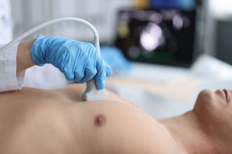
2D Echo & 4D Echo Services for Advanced Heart Evaluation in Amravati, Maharashtra
2D Echo and 4D Echo are non-invasive cardiac imaging tests that use ultrasound to assess the heart’s structure, movement and function. Dr. Niraj uses echocardiographic evaluation as part of appropriate cardiac assessment in Amravati, Maharashtra, helping assess heart function, valves and other structural abnormalities.
What it is
2D Echocardiography (2D Echo) uses ultrasound waves to create real-time images of the heart.
It can help evaluate:
- Heart chambers
- Heart muscle movement
- Pumping function
- Heart valves
- Blood flow
- Structural abnormalities
- Pericardial fluid
4D Echo provides advanced three-dimensional imaging with movement over time, where the technology and clinical service are available and appropriate.
2D Echo
A 2D Echo provides dynamic images of the beating heart.
It may be used to assess:
- Left ventricular function
- Right ventricular function
- Heart chamber size
- Valve structure and function
- Regional wall-motion abnormalities
- Pericardial effusion
- Structural heart abnormalities
The test is generally painless and does not involve radiation.
4D Echo
4D Echo provides three-dimensional visualization of cardiac structures with motion over time.
It can provide additional anatomical information in selected clinical situations, particularly when detailed visualization of cardiac structures or valves is required.
The usefulness of 4D imaging depends on the patient’s condition, image quality and the echocardiographic technology available.
Who May Need an Echo
An echocardiogram may be recommended for patients with:
- Breathlessness
- Chest discomfort
- Palpitations
- Heart murmurs
- Swelling of the legs
- Unexplained fatigue
- Suspected heart failure
- Known heart valve disease
- Cardiomyopathy
- Coronary artery disease
- Certain congenital heart conditions
It may also be used for monitoring previously diagnosed cardiac conditions.
Causes and Conditions Assessed
Echocardiography can help assess conditions affecting:
- Heart muscle
- Heart valves
- Heart chambers
- Heart function
- Structural anatomy
- Pericardium
It may contribute to the evaluation of heart failure, cardiomyopathy, valve disease, structural heart disease and other cardiac conditions.
Risk Factors
The need for echocardiography may be higher in patients with cardiovascular risk factors or a history of:
- High blood pressure
- Diabetes
- Coronary artery disease
- Previous heart attack
- Heart failure
- Heart valve disease
- Cardiomyopathy
- Congenital heart disease
- Abnormal heart rhythms
An Echo may also be recommended based on symptoms or findings during a cardiac examination.
Symptoms That May Require Evaluation
Symptoms that may lead to an echocardiogram include:
- Breathlessness
- Chest discomfort
- Palpitations
- Fatigue
- Dizziness
- Fainting
- Reduced exercise tolerance
- Leg or ankle swelling
The need for an Echo depends on the clinical assessment.
Red-Flag Symptoms
An Echo is not a substitute for emergency treatment.
Seek immediate emergency medical attention for:
- Severe or persistent chest pain
- Severe breathlessness
- Fainting or loss of consciousness
- Sudden severe weakness
- New neurological symptoms
- Symptoms suggestive of a heart attack
What Happens During a 2D Echo
During a standard 2D Echo:
- The patient generally lies on an examination bed.
- Small electrodes may be used for ECG monitoring.
- Ultrasound gel is applied to the chest.
- A handheld ultrasound probe is placed at different positions on the chest.
- Images and measurements of the heart are recorded.
- The findings are assessed as part of the cardiac evaluation.
The procedure is generally non-invasive and does not involve radiation.
What Happens During a 4D Echo
A 4D Echo uses specialized ultrasound technology to obtain three-dimensional images with movement over time.
The examination is performed using an ultrasound probe, with image acquisition depending on the cardiac structure being evaluated and the available technology.
Preparation
For many standard echocardiograms, extensive preparation is not required.
Patients should follow any specific instructions provided before the examination.
If the Echo is being performed as part of a broader cardiac evaluation, additional investigations or preparation may be recommended depending on the clinical situation.
Recovery
A standard 2D Echo or 4D Echo generally requires no significant recovery period.
Patients can typically resume normal activities after the examination unless another procedure or test has been performed at the same appointment.
Potential Limitations
Echocardiographic image quality can vary between patients.
Factors that may affect imaging include:
- Body habitus
- Lung interference
- Patient positioning
- Movement during the examination
- Image quality
- The specific cardiac structure being assessed
Additional cardiac imaging may sometimes be required when an Echo does not provide sufficient information.
2D Echo vs 4D Echo
2D Echo provides two-dimensional real-time images and is widely used for routine assessment of heart structure and function.
4D Echo provides three-dimensional visualization with movement over time and may offer additional anatomical information in selected cases.
The appropriate imaging method depends on the clinical question and available technology.
Diagnosis
Echocardiography may contribute to the assessment of:
- Heart failure
- Cardiomyopathy
- Valve disease
- Coronary artery disease
- Structural heart disease
- Congenital heart disease
- Pericardial conditions
- Cardiac function
The Echo findings are interpreted together with symptoms, medical history and other investigations.
When 2D or 4D Echo May Be Required
An echocardiogram may be recommended when a doctor needs to evaluate:
- Heart pumping function
- Valve function
- Heart chamber size
- Heart muscle movement
- Structural abnormalities
- Causes of breathlessness
- Suspected heart failure
- Known cardiac disease
- Changes in previously diagnosed heart conditions
4D imaging may be considered when additional three-dimensional anatomical information is clinically useful and the technology is available.
Medical Treatment
2D Echo and 4D Echo are diagnostic investigations rather than treatments.
The results may help guide treatment for conditions such as:
- Heart failure
- Cardiomyopathy
- Valve disease
- Coronary artery disease
- Structural heart disease
- Arrhythmias
Treatment depends on the diagnosis established through the overall cardiac evaluation.
Treatment Alternatives
Depending on the clinical question, other investigations may include:
- ECG
- Holter monitoring
- Stress testing
- CT coronary angiography
- Cardiac CT
- Cardiac MRI
- Coronary angiography
- Other specialized cardiac imaging
The appropriate investigation depends on the patient’s symptoms and suspected condition.
Related Procedures
Related cardiac services include:
- Advanced Cardiac Imaging
- Coronary Angiography
- Coronary Physiology
- Intravascular Imaging
- Structural Heart Programme
- TAVR / TAVI
- Mitral Valve Interventions
- Tricuspid Interventions
- Heart Failure Management
- Electrophysiology & Arrhythmia
FAQs
What is a 2D Echo?
A 2D Echo is an ultrasound examination that creates real-time two-dimensional images of the heart to assess its structure and function.
What is a 4D Echo?
A 4D Echo provides three-dimensional visualization of cardiac structures with movement over time using specialized ultrasound technology.
Is 2D Echo painful?
A standard 2D Echo is generally painless and non-invasive. Some pressure may be applied with the ultrasound probe while obtaining images.
Does an Echo use radiation?
No. Standard echocardiography uses ultrasound waves and does not use ionizing radiation.
Why might I need a 2D Echo?
It may be recommended to evaluate heart function, valves, chambers, structural abnormalities or symptoms such as breathlessness, chest discomfort or palpitations.
What is the difference between 2D Echo and 4D Echo?
2D Echo produces two-dimensional real-time images, while 4D Echo can provide three-dimensional visualization with movement over time in suitable clinical situations.
How long does an Echo take?
The duration can vary depending on the type of examination, the images required and the patient’s clinical condition.
Do I need special preparation for an Echo?
Many standard echocardiograms require little or no special preparation. Follow the specific instructions provided for your examination.
Can an Echo detect heart failure?
An echocardiogram can provide important information about heart structure and pumping function that contributes to the assessment of heart failure.
Can an Echo detect valve problems?
Yes. Echocardiography can evaluate the structure and function of the heart valves and help assess abnormalities.
When should I consult Dr. Niraj in Amravati?
If you have breathlessness, palpitations, chest discomfort, a heart murmur, swelling, reduced exercise tolerance or a known heart condition, Dr. Niraj can determine whether echocardiographic evaluation is appropriate.
Book an Appointment with Dr. Niraj for 2D Echo & 4D Echo Evaluation in Amravati
If you have breathlessness, palpitations, suspected heart disease, valve problems, heart failure or another cardiac concern, Dr. Niraj can assess your symptoms and determine whether 2D Echo, 4D Echo or another cardiac investigation is appropriate.